


Osteoarthritis of the ankle joint is a chronic disease that affects the articular cartilage, and subsequently other structures of the joint (capsule, synovium, bones, ligaments). It has a degenerative-dystrophic character. It presents with pain and limitation of movements, followed by progressive impairment of support and walking functions. Diagnosis is based on symptoms, examination and radiography. Treatment is usually conservative, using anti-inflammatory drugs, chondroprotectors, and glucocorticoids, and prescribing exercise and physical therapy. In severe cases, sanitary arthroscopy, arthrodesis or endoprosthesis is performed.
Main information
Osteoarthritis of the ankle joint is a disease in which the joint cartilage and surrounding tissues are gradually destroyed. The disease is based on degenerative-dystrophic processes, inflammation in the joint is secondary. Arthrosis has a chronic undulating course with alternating remissions and exacerbations and gradually progresses. Women and men suffer equally often. The likelihood of development increases sharply with age. At the same time, experts note that the disease is "getting younger" - every third case of ankle arthrosis is currently found in people under 45 years of age.
reasons
Primary arthrosis occurs for no apparent reason. Secondary damage to the ankle joint develops under the influence of some adverse factors. In both cases, the basis is a violation of the metabolic processes in the cartilage tissue. The main causes and predisposing factors for the formation of secondary arthrosis of the ankle joint are:
- major intra- and peri-articular injuries (fractures of the talus, fractures of the ankle, lacerations and ruptures of ligaments);
- ankle surgery;
- excessive load: too intense sports, long walking or constant standing due to working conditions;
- wearing shoes with heels, overweight, permanent microtraumas;
- diseases and conditions related to metabolic disorders (diabetes mellitus, gout, pseudogout, estrogen deficiency in postmenopause);
- rheumatic diseases (SLE, rheumatoid arthritis);
- osteochondrosis of the lumbar spine, intervertebral hernia and other conditions that are accompanied by pinching of nerves and disruption of the muscular system of the foot and leg.
Less often, the cause of arthrosis is non-specific purulent arthritis, arthritis due to specific infections (tuberculosis, syphilis) and congenital developmental anomalies. Unfavorable environmental conditions and hereditary predisposition play a certain role in the development of arthrosis.
Pathogenesis
Usually, the articular surfaces are smooth, elastic, smoothly slide relative to each other during movements and provide effective shock absorption during loading. As a result of mechanical damage (trauma) or metabolic disorders, cartilage loses its smoothness, becomes rough and inelastic. The cartilage "rubs" during movements and injures each other, which leads to worsening of pathological changes.
Due to insufficient cushioning, the excess load is transferred to the underlying bone structure and degenerative-dystrophic disorders develop in it: the bone is deformed and grows along the edges of the joint area. Due to secondary trauma and disruption of the normal biomechanics of the joint, not only the cartilage and bones suffer, but also the surrounding tissues.
The joint capsule and synovial membrane are thickened and foci of fibrous degeneration are formed in the ligaments and periarticular muscles. The ability of the joint to participate in movements and withstand loads is reduced. Instability occurs and pain progresses. In severe cases, the joint surfaces are destroyed, the supporting function of the limb is impaired and movements become impossible.
Symptoms
Initially, rapid fatigue and slight pain in the ankle joint are detected after a significant load. Subsequently, the pain syndrome becomes more intense, its character and time of occurrence change. Distinctive features of arthrosis pain are:
- Initial pain. They appear after a state of rest and then gradually disappear with movement.
- Dependence on load. There is increased pain during physical exertion (standing, walking) and rapid fatigue of the joint.
- Night pain. They usually appear in the morning.
The condition changes in waves, during exacerbations the symptoms are more pronounced, in the phase of remission they first disappear, then become less intense. There is a gradual progression of symptoms over several years or decades. Along with pain, the following manifestations are determined:
- A crunching, creaking or clicking sound may be heard during movement.
- During an exacerbation, the periarticular area sometimes swells and becomes red.
- Due to instability of the joint, the patient often twists the leg, causing sprains and tears in the ligaments.
- Stiffness and limitation of movements are noted.
Complications
During an exacerbation, reactive synovitis may occur, accompanied by fluid accumulation in the joint. In the later stages, a pronounced deformity is revealed. Movements are sharply limited and contractures develop. Support becomes difficult, when moving patients are forced to use crutches or a cane. There is a reduction or loss of ability to work.
Diagnosis
The diagnosis of arthrosis of the ankle joint is made by an orthopedic doctor based on a study, data from an external examination and results of additional tests. When examining in the initial stages, there may be no changes, but later deformations, limitation of movements and pain on palpation are detected. Leading importance is given to visualization techniques:
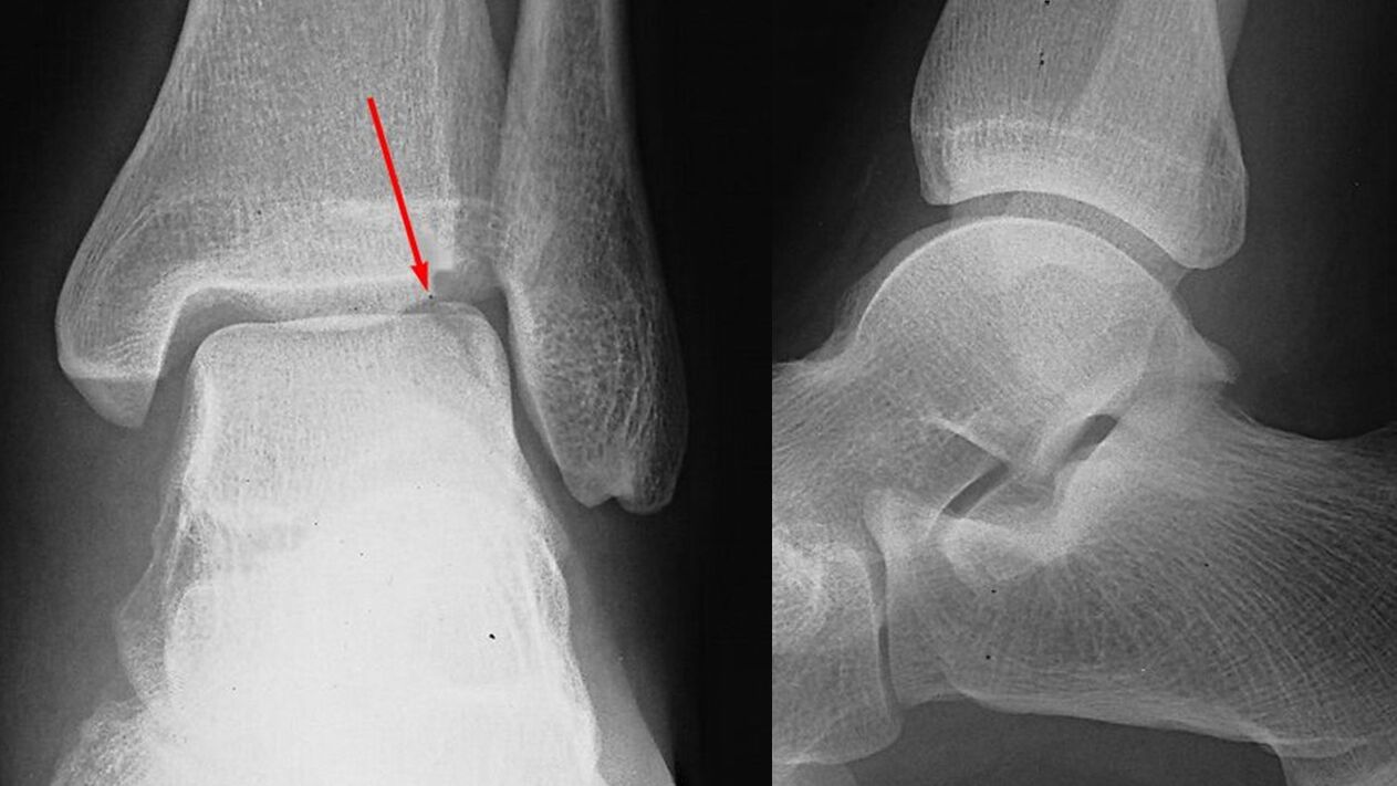
- X-ray of the ankle joint. It plays a decisive role in establishing the diagnosis and determining the degree of arthrosis. The pathology is manifested by narrowing of the joint gap, proliferation of the edges of the joint surfaces (osteophytes). At a later stage, cystic formations and osteosclerosis of the subchondral (located under the cartilage) area of the bone are found.
- Tomographic studies. Used when specified. In difficult cases, for a more accurate assessment of the condition of the bone structures, the patient is additionally sent for a computed tomography, and for examination of the soft tissues - for an MRI of the ankle joint.
Laboratory tests are unchanged. If necessary, to establish the cause of arthrosis and differential diagnosis with other diseases, consultations with related specialists are prescribed: neurologist, rheumatologist, endocrinologist.
Treatment of arthrosis of the ankle
The treatment of the pathology is long-term and complex. Patients are usually seen by an orthopedic surgeon on an outpatient basis. In the period of exacerbation, hospitalization in the department of traumatology and orthopedics is possible. The most important role in slowing down the progression of arthrosis is played by lifestyle and the correct physical activity regime, which is why the patient is given recommendations to reduce weight and optimize the load on the leg.
Drug therapy
It is chosen individually, taking into account the stage of arthrosis, the severity of symptoms and concomitant diseases. Includes general and local agents. The following groups of drugs are used:
- General NSAIDs. Tablet forms are usually used. Medicines have a negative effect on the gastric mucosa, so "gentle" medicines are preferred for gastrointestinal diseases.
- Local NSAIDs. It is recommended both in the period of exacerbation and in the phase of remission. It can be prescribed as an alternative if side effects occur from the tablet forms. It is available in the form of ointments and gels.
- Chondroprotectors. Substances that help normalize metabolic processes in cartilage tissue. They are used in the form of creams, gels and preparations for intra-articular administration. Use medications containing glucosamine and collagen hydrolyzate.
- Hormonal agents. For severe pain that cannot be relieved by drugs, intra-articular corticosteroids are administered no more than 4 times a year.
- Metabolic stimulants. To improve local blood circulation and activate tissue metabolism, nicotinic acid is prescribed.
Physiotherapy treatment
The patient is prescribed a complex of physiotherapy, developed taking into account the manifestations and stage of the disease. The patient is referred to physical therapy. In the treatment of arthrosis, massage and UHF are used. In addition, in the treatment of pathology they use:
- laser therapy;
- heat treatments;
- medicinal electrophoresis and ultraphonophoresis.
surgery
It is shown in the later stages of the disease, when conservative therapy is ineffective, severe pain syndrome, deterioration of the patients' quality of life or limited work capacity. The operations are performed in hospital conditions and are open and minimally invasive:
- Arthroscopic interventions. If there is significant cartilage destruction, arthroscopic chondroplasty is performed. Sanitary arthroscopy (removal of formations that interfere with movement) is usually performed for severe pain in stage 2 arthrosis. The effect lasts for several years.
- Arthrodesis of the ankle joint. It is performed with significant destruction of the joint surfaces, involves removing the joint and "fusion" of the bones of the foot and lower leg. Provides restoration of the limb's support function in case of loss of joint mobility.
- Endoprosthesis of the ankle joint. It is performed in advanced arthrosis. It involves removing the destroyed articular surfaces of the bones and replacing them with plastic, ceramic or metal prostheses. The movements are completely restored, the service life of the prosthesis is 20-25 years.
Forecast
The changes in the joint are irreversible, but the slow progression of arthrosis, the timely initiation of treatment and compliance with the recommendations of an orthopedic traumatologist in most cases allow maintaining work capacity and a high quality of life for decades after the onset. from the first symptoms. With a rapid increase in pathological changes, endoprosthesis allows to avoid damage.
Prevention
Preventive measures include reducing the level of injuries, especially in winter, during periods of ice. If you are overweight, it is necessary to take measures to reduce body weight to reduce the load on the joint. You should maintain a regime of moderate physical activity, avoid overloads and microtraumas, promptly treat diseases that can provoke the development of arthrosis of the ankle joint.